Background
One important symptom of water stress on plants is the formation and propagation of air embolisms within the xylem – the interconnected tubular network of cells that transport water from the roots to the rest of the plant. The invasion of air by cavitation progressively reduces the plant’s ability to transport water, which impacts plant productivity and eventually leads to death.
The relationship between embolism formation and the degree of water stress experienced by the plant is known as xylem vulnerability, and is a key metric used to identify and compare drought tolerance between species and individuals. Knowing these differences, and understanding tolerances to levels of water availability, is critical in an age where the limits of agricultural and natural plant communities are being tested by rapid environmental change.
Current methods of assessing xylem vulnerability typically involve techniques where the restriction in water movement due to embolism is measured in water pushed or pulled through the xylem of excised stems and leaves. Although there is general consensus among researchers that in most cases these approaches can provide valid assessments of vulnerability, there are concerns and evidence of methodological artefacts that arise from the nature of the sample preparation, a procedure which is unavoidably invasive and damaging.
A more direct and non-invasive technique called X-ray micro-tomography, or microCT, is fast becoming the gold standard, and can produce stunning high-resolution 3D images of the xylem, in which it is possible to identify embolism at the cellular level. However, the technique is expensive, time-consuming, and access to the technology limited to a few facilities, which are typically high in demand. In addition, only small sections of xylem can be imaged at at time, which limits any extrapolation of results to the plant as a whole, and repeated imaging causes sample degradation.
A new technique
In 2015 researchers at the University of Tasmania, in collaboration with the LiPhy national scientific research centre in Grenoble in France, realised that there was a subtle, but visible, change in the brightness of non-embolised (water-filled) xylem after it transitions to become embolised (air-filled). From this realisation was born a new and exciting, and relatively inexpensive and simple optical technique for identifying embolism formation and assessing vulnerability.
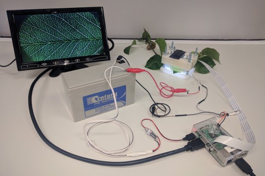
Uninterrupted power supply for the RaspberryPi setup using a battery and power converter
Read article All articles Play
Play
Eucryphia lucida leaf 2
Captured by Jen Peters from Western Sydney University visiting the Brodribb Lab in Hobart.