Capture
If you haven’t done so already take a look at the overview to get an understanding of the overall procedure, the background to the technique, and information about the project in general.
The first stage of the process is to capture images of the xylem over time. This can be achieved using one of four methods:
- A digital camera mounted on a microscope
- A digital camera with a good quality macro lens
- A desktop scanner
- A custom built OpenSourceOV clamp (OSOV)
- A commercial “Cavicam” – see https://cavicam.co (or https://cavicams.com)for more information
Instructions for capturing sequences using the first two methods are not currently available (although you can help write them) because of the large number of options and configurations available for digital cameras, microscopes, lenses, and camera-control software. In the meantime it shouldn’t be too difficult to get something setup, the key components required are:
- Suitably bright illumination through the leaf or above the stem
- Good quality lens. The magnification will depend on the desired analysis – to capture whole leaf events a standard macro lens will suffice but for more detailed analysis a lens with 20x and greater magnification is required.
- Time-lapse function to control the timing and duration of captures
Some things to consider when putting together your own setup:
- Most cameras and camera-control software have a time-lapse function but beware that some have a limit to the length of capture e.g. a day. Check the manual before purchase.
- Captured images should always be saved in a file format that is lossless i.e. has not undergone any compression to reduce file size, such as jpg or gif. Lossless image formats include raw, tiff, png, and bmp. Compressed formats introduce artefacts as a result of the compression process.
Regardless of the capture method the processing instructions will be the same. Once you have your sequence of captures head over to process for the next stage of the procedure.
Instructions for capturing images using a desktop scanner and OSOV clamp can be found below:
Resources
Instructions for building the OSOV clamp
http://github.com/OpenSourceOV/clamp-build-instructions
Capturing images using the OSOV clamp
http://github.com/OpenSourceOV/clamp-image-capture-instructions
Capturing images using a desktop scanner
http://github.com/OpenSourceOV/scanner-image-capture-instructions
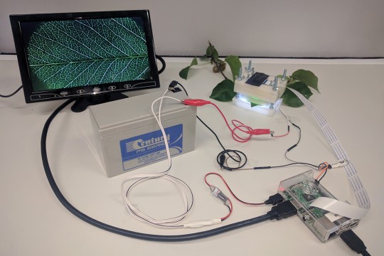
Uninterrupted power supply for the RaspberryPi setup using a battery and power converter
Read article All articles Play
Play
Eucryphia lucida leaf 2
Captured by Jen Peters from Western Sydney University visiting the Brodribb Lab in Hobart.