An integrated device
A setup that employs a camera with large light sensors and a good quality macro lens will produce excellent low-noise images of embolism in action, but the equipment will be expensive, bulky, and not easily mobile. The setup also requires construction of an appropriate stage with suitable lighting so the sample can be fixed in position and sufficiently illuminated.
It is clear that the evolution of this setup is to incorporate all of these components – camera, lens, illumination, stage, processing unit – into a single integrated device that also addresses issues of cost and mobility.
Happily this perfectly coincides with a market exploding with cheap programmable micro-controllers and computers, such as the Arduino and Raspberry Pi, to which can be connected small high resolution cameras. Add to that a meteoric rise in 3D printing technology, which has reached such maturity that is now remarkably easy for anyone to design and fabricate precise and durable parts, in a range of materials, by yourself, or using a growing service industry of online 3D printers.
But what about the problem of noise? Indeed, miniaturised cameras have smaller light sensors, which means more noise, and this is certainly a challenge for this type of device. However, noise removal algorithms can be remarkably effective, and there are other ways of amplifying the signal; by providing more light to each sensor (increasing illumination) and increasing the signal over more sensors (increasing magnification).
The first integrated device was developed by researchers at the University of Tasmania in 2016. The device – ‘the clamp’ – utilises a Raspberry Pi micro-computer for storage and control, a small high-resolution camera for image capture, super-bright LEDs for illumination, and a 10x smart-phone macro lens for magnification, all contained within a 3D-printed assembly that incorporates a stage for fixing the sample in position.
 Play
Play
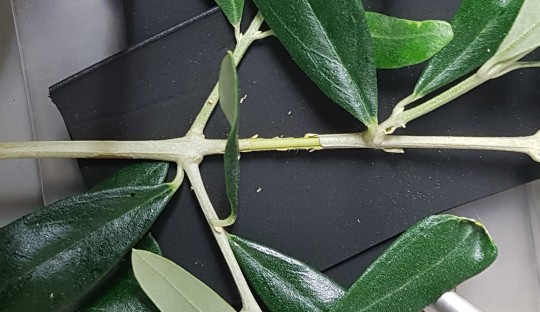
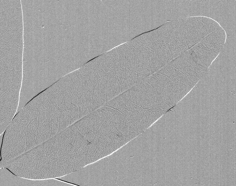
Eucryphia lucida leaf 2
Captured by Jen Peters from Western Sydney University visiting the Brodribb Lab in Hobart.